IND
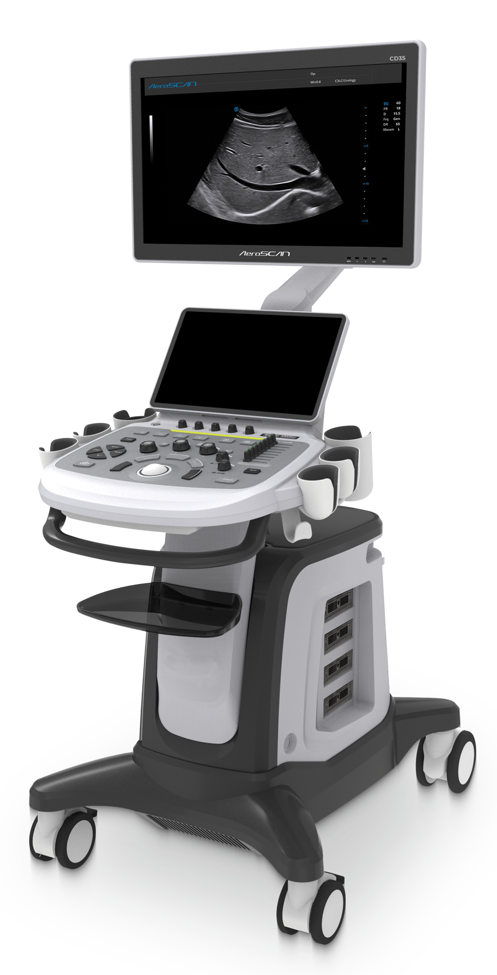
Aeroscan CD35 is an Advanced Cart Based Color Doppler Ultrasound System with 5Q Probe Technology enhance High Quality B-mode resolutions and increase the stability of probe performance, the homogeneity of image resolution and the High Sensitive on Color Doppler, CD35 innovatively designed for fast user experience with Limited Keys
23 Inch Big HD Monitor
13.3 Inch Touch Screen
4 Pinless Probe Connector
Rotatable Control Panel
with Elevation
Advanced user interface with Smooth workflow
Output- 4 USB, Dicom 3.0,
VGA, S-Video
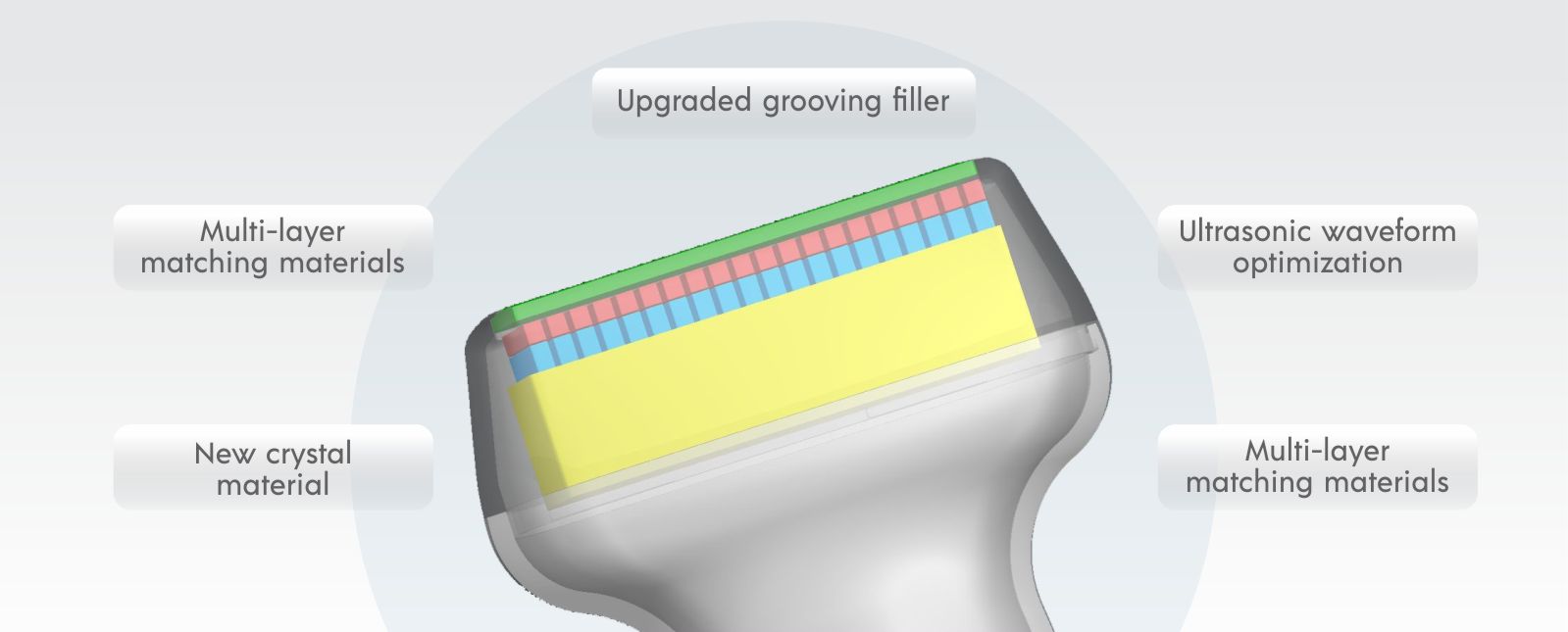
5Q Probe Technology
5Q probe technology refers to five technologies that improve the image quality, including high performance PZT crystal materials, crystal cutting technology, element spacing filler, multiple matching layer technology and optimized sound field wave. All these technologies integrated to increase the probe bandwidth and enhance it's sensitivity as well as resolution.
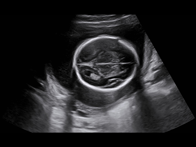
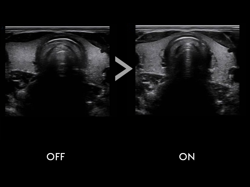
Fusion THI 2.0
Fusion THI 2.0
Based on the new generation of invention tissue Harmonic, Fusion THI 2.0 integrated fundamental Wave and harmonic wave in the far field. With greatly enhanced tissue harmonic wave, it delivers penetration improved image by effectively increasing image resolution and tissue contrast.
Xbeam
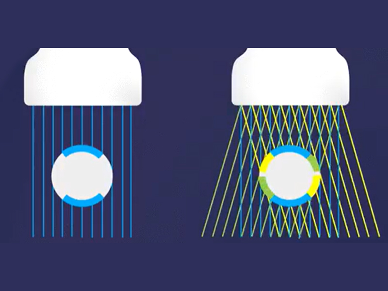
Xbeam
The upgraded Xbeam is a new processing mode based at the front end of ultrasound beamforming. With steerable beam angle and the high frame rate, Aeroscan CD35 effectively increases the spatial resolution and reduces tissue shadow. Xbeam applies to most kinds of probes.
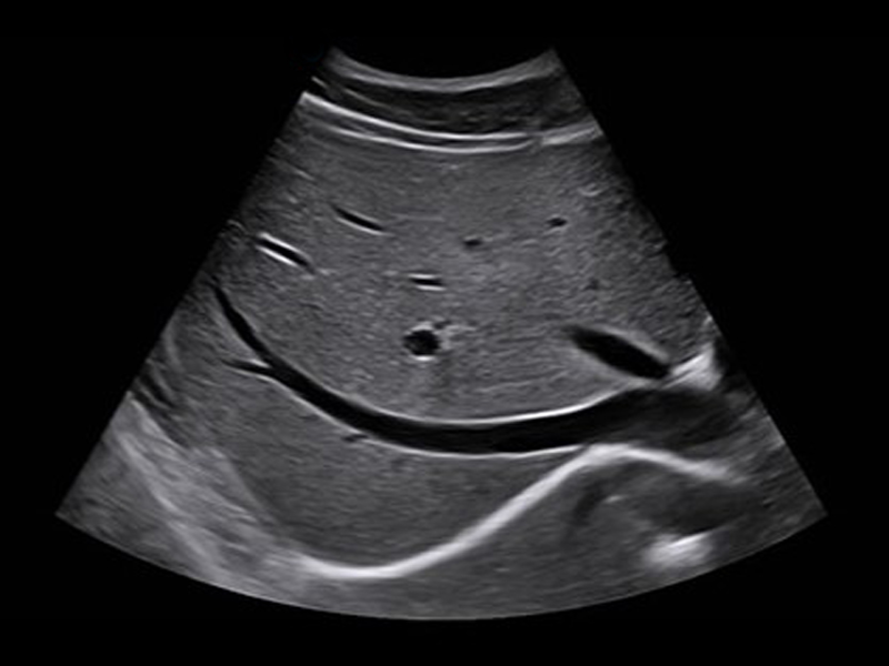
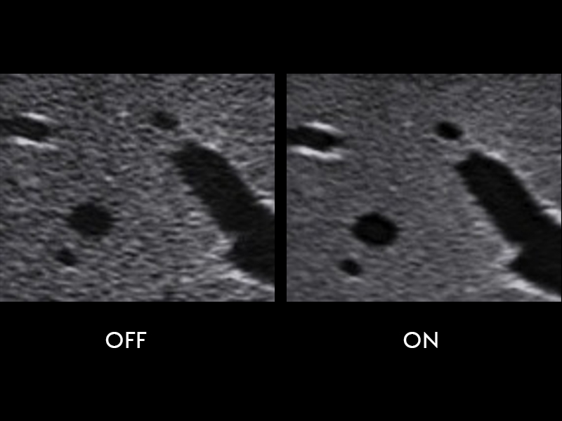
Balance Echo Compensation
Balance Echo Compensation
The upgraded AeroACAN CD35 transplant Balanced Echo Compensation technology from the High - End platform. It effectively compensate the weak echo signals and restrains the hyper echo signals according to the grey scale distribution and delivers much more uniform image.
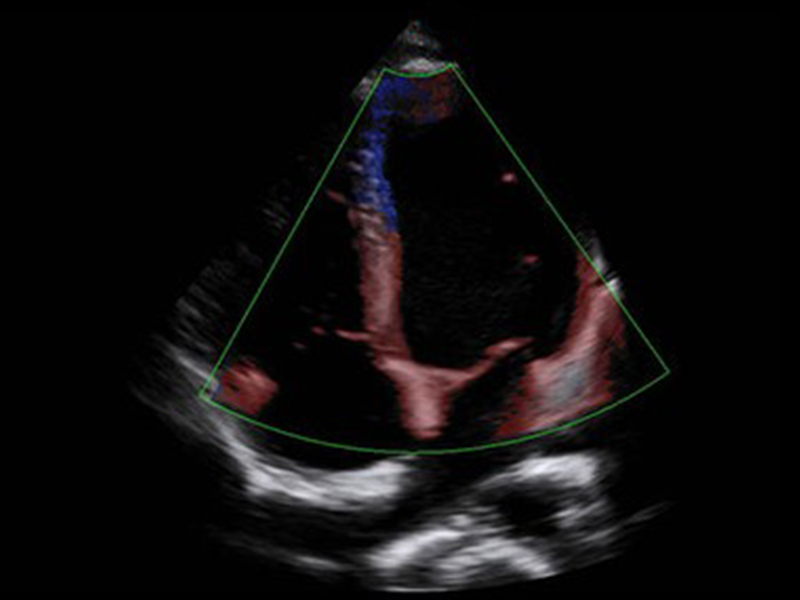
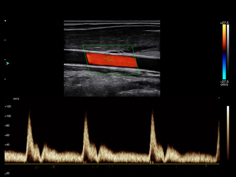
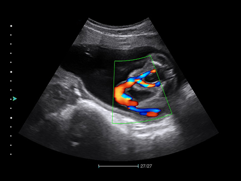
VS Flow
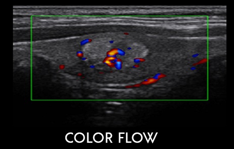
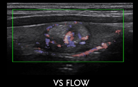
VS Flow
VS flow is a high spatial resolution Doppler flow imaging. 30% more side signal is collected to enhance the whole flow sensitivity especially the slow velocity flow.
Elastography
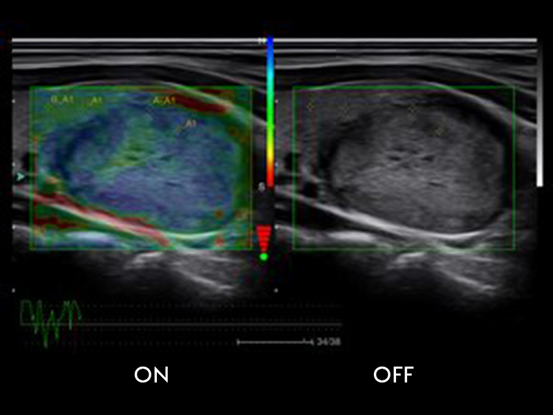
Elastography
Elastography offers a real-time tissue stiffness assessment to detect potential abnormalities within normal tissue such as thyroid, breast, superficial tissue, etc. Qualitative measurement supports effectively distinguish between benign and malignant solid tumors.
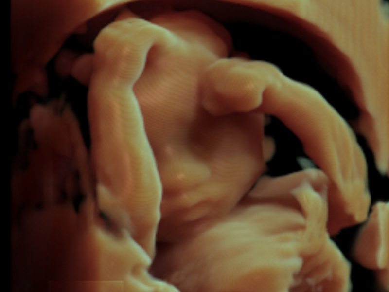
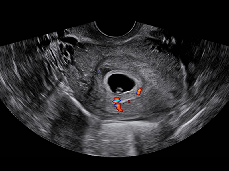
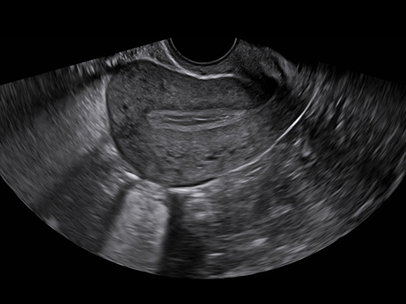
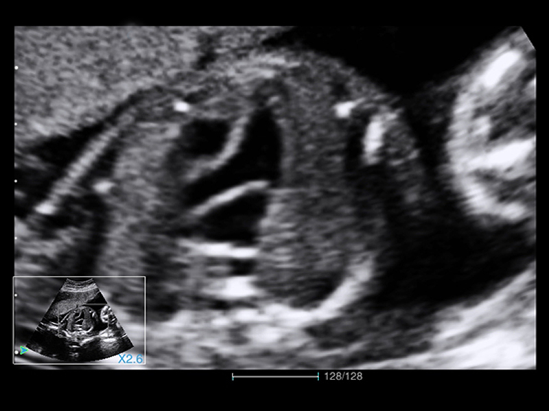
Lumi 4D
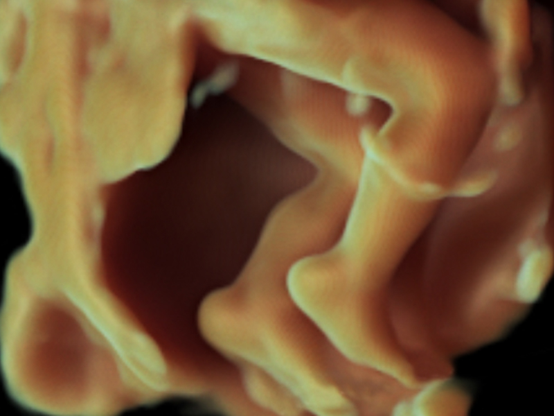
Lumi 4D
Utilizing light source with adjustable angle, it presents a vivid fetal face and a much stronger three-dimensional sense.: the system supports abdominal and vaginal volume probes for 3D / 4D imaging in OB / GYN applications, providing more detailed and complete volume information to help observe and diagnose fetal health and uterine health.
Technical specifications
Imaging Mode
B, B/B, 4B, B/M, M, B/D, THI, Color, Power & Directional Power, PW/ CW Doppler, Duplex, Triplex, Color M, Anatomical M Mode, TDI, Panoramic Imaging, 4D Imaging, Auto IMT Calculations.
Probe Connector
4 live probe connectors.
Image Magnification
Full Screen Zoom, HD Zoom.
Printer
Direct Printer Support
Clinical Application
Radiology, Vascular, Thyroid, OBS/GYN, MSK, Adult/Peadiatric Cardiac, Urology, Pediatrics, Neonatal Head, Small parts, Transcranial etc.
Probes
Convex / Linear / Micro Convex /Phased Array/Trans Vaginal / Transrectal / Volume Convex / Volume TV Probe and More.,
Advance Features
Trapezoid Imaging, Nanoview, Auto-fit, XBeam, MFI, Free Hand 3D, IMT Auto Measurement, Smarchive, ECG Module, VS Flow, Panoramic, 4D Pro + 4D Lite, CW, Anatomic M, Color M, TDI and More.,
Gallery