IND
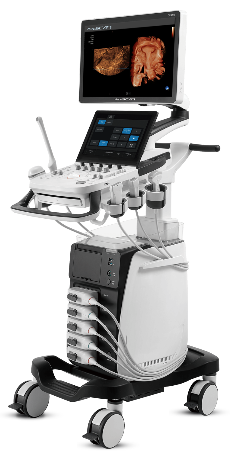
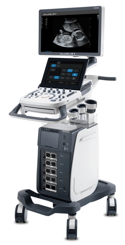
Aeroscan CD40 Premium segment systems provides intelligent workflow, Reliability, Outstanding Performance, Premium image Quality, Highly sensitive Color Doppler, Durability Hardware & Software, CD40 provides quick and fast diagnostic confidence to the doctors, The Intelligent Workflow and User Friendly Control Panel providing efficiency and remarkable user day to day experience

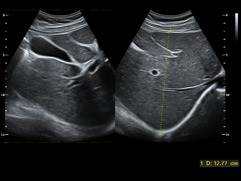
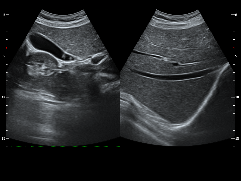
Radiology

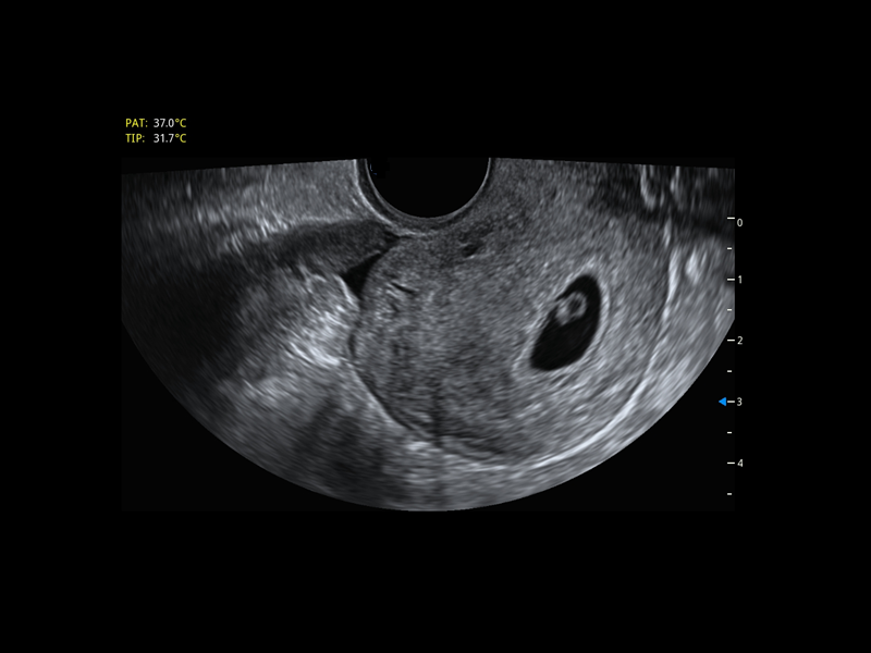
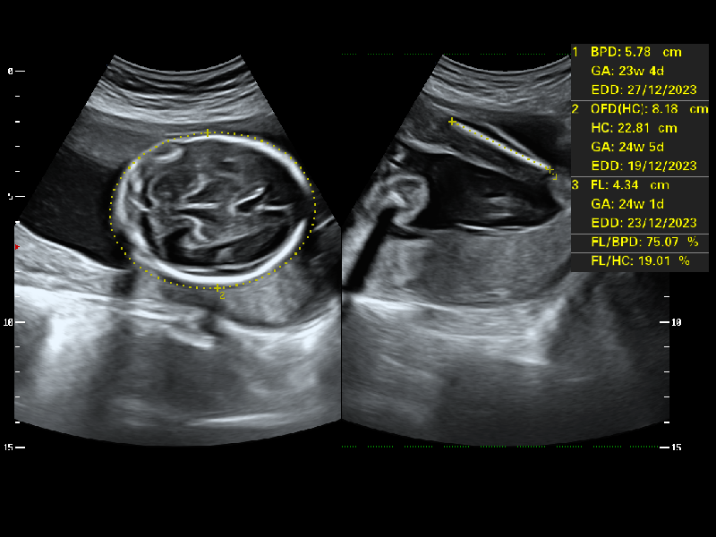
OB-GYN

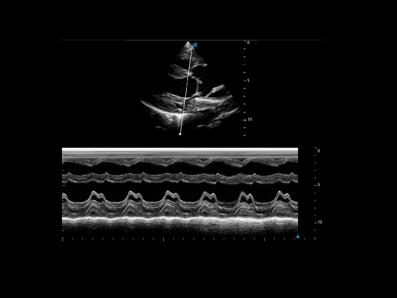
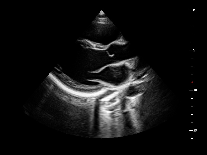
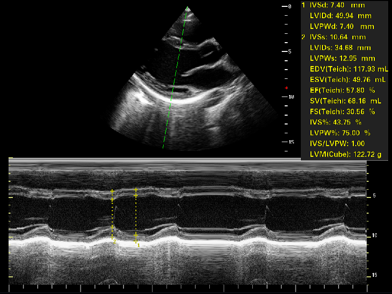
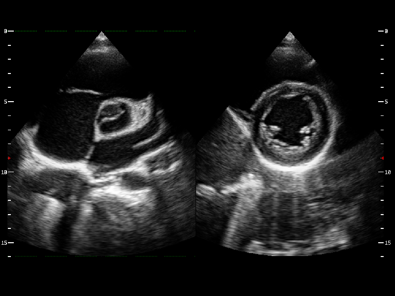
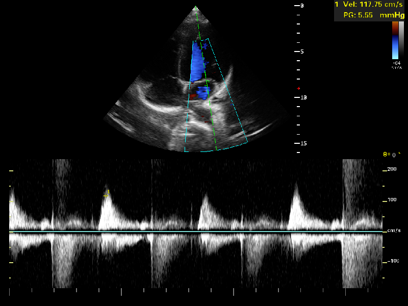
Cardiology

Point of Care
Advanced Technologies for
Unexceptionable Images
μ-Scan+ - Advance Speckle Reduction
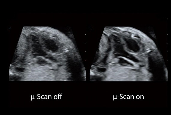
Advance μ-Scan+
A Latest generation μ-Scan+, Play major role in B- Mode and 3D/4D modes, is more delicately engineered to distinguish tissue and artifacts, Improves the visibility of organs and Lesions in B Mode & Enhanced the B-Mode Resolution . In the meantime control the Speckle noise , improving image uniformity and enhance border continuity.
Spatial Compound Imaging
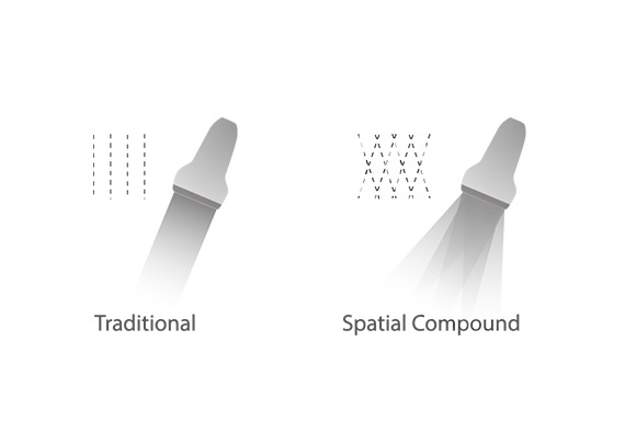
Spatial Compound Imaging
Spatial Compound Imaging utilizes several lines of sight for optimal contrast resolution, speckle reduction and border detection, with which CD40 is ideal for superficial and abdominal imaging with better clarity and improved continuity of structures.
Advance Dynamic Color & HPRF
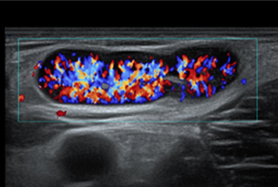
Advance Dynamic Color & HPRF
Effectively Improve the dynamics Resolution and Increase the resolution of blood Flow.
Clinical Benefit:
Low & Slow Blood Flow Detection for better and accurate vessel profile , High Spatial resolution for less overfilling & Greater Color Doppler Sensitivity.
Advance Feature
-

21.5 inch LED monitor
-

-

Multi-stage temperature control gel warmer
-

13.3- Inch high sensitivity touch screen adjusts to accommodate user viewing preference in any scanning environment
-

5 active transducers sockets provides wide range of clinical applications
-

Sliding keyboard
-

Large capacity detachable battery
Advance Feature and 4d Volume
Auto Face
3D imaging of the fetal face has important implications for the diagnosis of facial malformations. Auto Face easily hides occlusions and artifacts such as umbilical cord, placenta, uterine wall and extremities to best display the fetal face in 3D.
S-Depth
S-Depth can automatically display the near-far relation from transducer to target, represented by a smart designed color coding. It helps doctors to judge the spatial relationship on real-time 3D images.
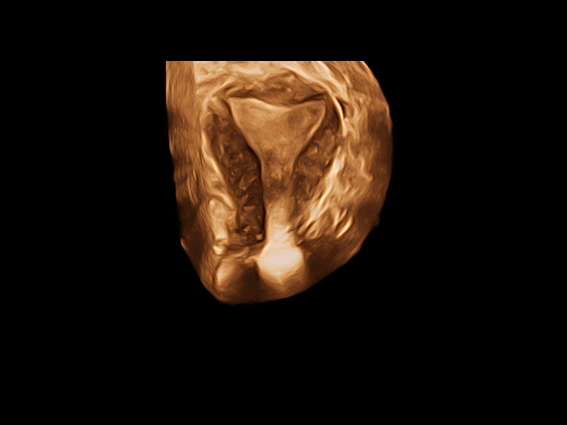
S-Live Silhouette
S-Live Silhouette is a unique transparent volume image for a more comprehensive internal and external view of the anatomy and provides more abundant diagnostic information for the clinical Applications.
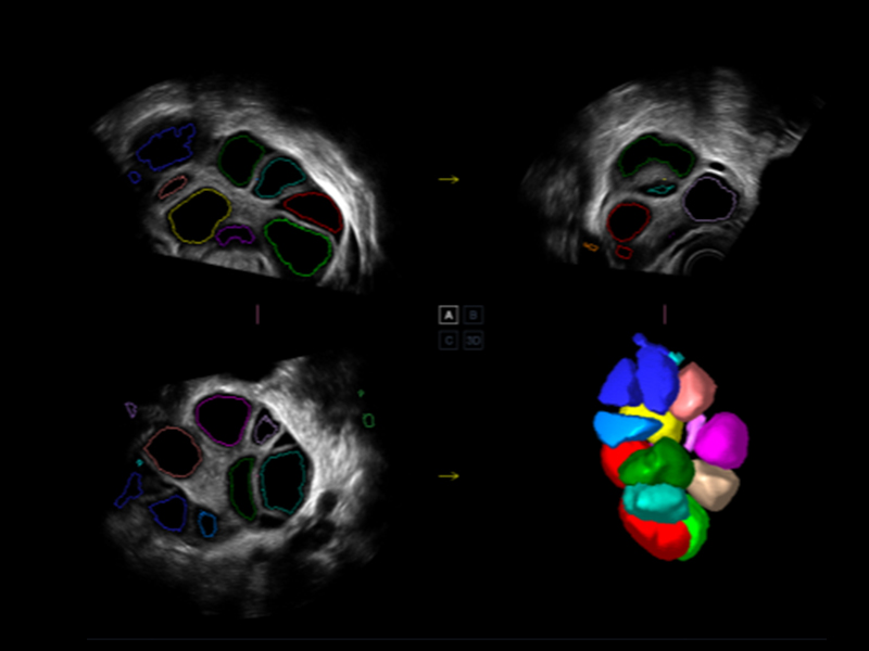
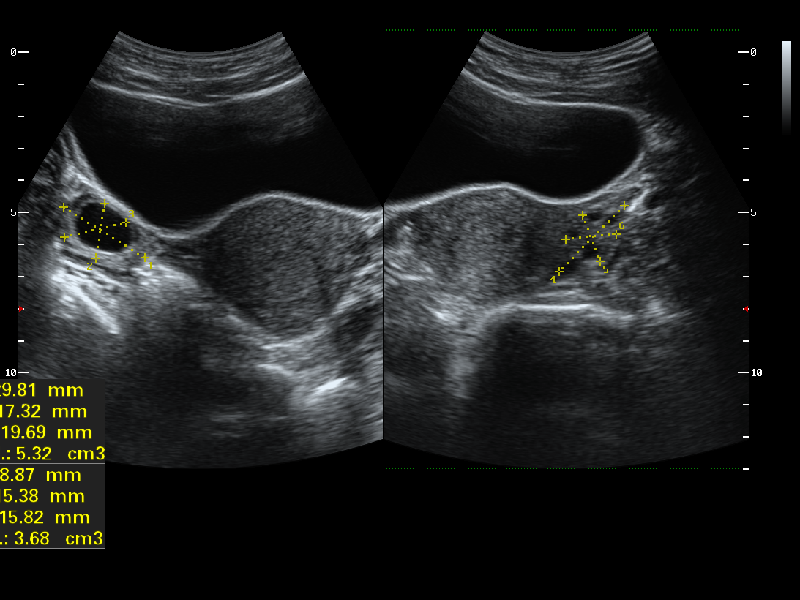
AVC Follicle
High Efficiency of follicle analysis is achieved by AVC Follicle, a Volume-data based automatic follicular calculation including the number and volume. Follicles are sorted by sizes in the results and rendered in different colors with numbers for better Visualization.
Contrast Imaging
The contrast agents provide a loud signal reflection, giving a more enhanced image of difficlut-to-view blood flow. Controls the acoustic pressure and provides promising image quality with a smaller agent dose.
C-xlasto Imaging
Strain elastography for evaluating tissue stiffness, professional semi-quantitative analysis with strain ratio indicating tissue elasticity and the system will rapidly calculate the Strain Ratio and display unusually hard or soft anomalies within the soft tissue.. Available in convex, transvaginal, Linear.
Auto NT
It allows to automatically measure the nuchal translucency. Auto NT Measurents allow a single click for the higher accuracy.
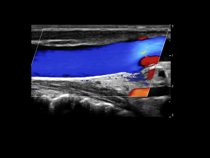
Auto IMT
Auto IMT is a smart tool to analyze a patient's potential risk of cardiovascular disease. By clicking a button, you can measure both the anterior and posterior intima-media thickness of the common carotid. This simple procedure enhances exam productivity as well as adds diagnostic value to the exam.
Auto EF
Recognize the Myocardial intimate During the Diastolica and Systolic Period and Calculate ejection Fraction Automatically.
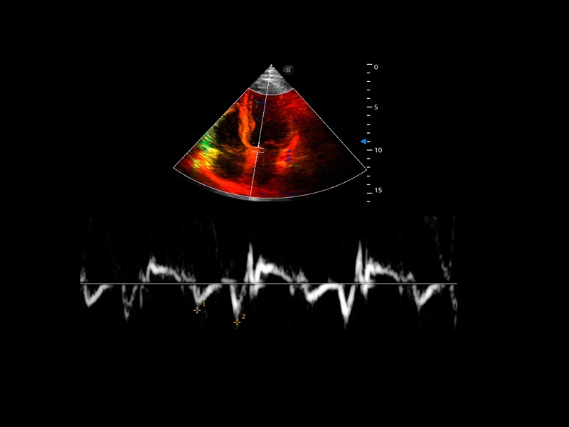
TDI
TDI uses myocardial Doppler frequency shifts to present an informative view of myocardial movement indicating velocity and direction.
Stress Echo
Stress echo is used to diagnose coronary heart disease, evaluate coronary reserve function and myocardial ischemia, and estimate myocardial viability, providing valuable diagnostic information for PCI&CABG.
Real-Time Color Panoramic
Combination of Color Flow and Real time panoramic, Visualizing the Blood Flow of an entire artery or vein is now very simple task with the Real Time Color Panoramic Accomplished in the real-time for Convenience of the user, any mistake can also be easily back tracked and corrected without interrupting the scan.
Vis-Needle
By emphasizing the visualization of the needle, it increases the safety and accuracy of biopsy procedures and other interventional procedures including nerve blocks and vascular accesses.
Advanced Transducer Technologies
1Mhz Low Frequency Convex transducer greatly improve the Piezoelectric crystal signal ratio, Acquire stunning Quality Images & New Generation μ-Scan + Provide High sensitivity & High Resolution for the Both near and far field, 200 degree Transvaginal Probe provides extraordinary image quality for gynaecology, Early Obstetrics scan, HD Linear Transducer achieve a Uniform image Quality, high sensitive acoustic spectrum in vascular, brest, MSK, Thyriod, Lower and upper limp, Etc and Aeroscan CD40 enhancing boundary improved visualization in 2D and Color Doppler in Echocardiogram with all premium Features loaded., CD40 Supports Ultras-wide Frequency bandwith Probes - Intra-operative, Paediatric, Neonatal, TEE, Transrectal, Volume Transducer & More.,
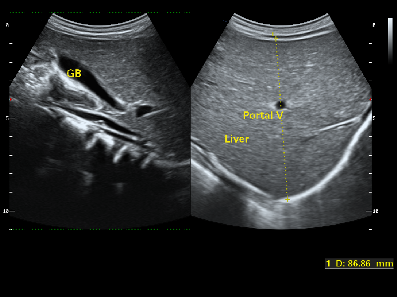
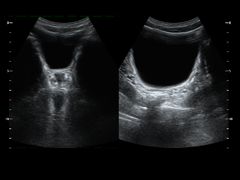
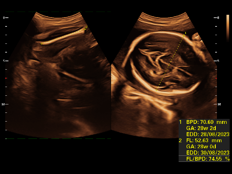
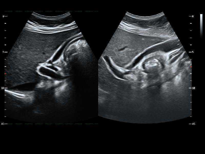
Gallery